Full Mouth and Intraoral X-ray Examination
Home > Oral Services > General Dentistry – Full Mouth and Intraoral X-ray Examination
Full Mouth and Intraoral X-ray Examination
In a typical routine dental examination, only the surface of the teeth or shallow gum health can be observed. In order to detect conditions such as root abnormalities, inflammation of the dental pulp, or internal tooth decay (cavities) within the teeth and jawbone, X-ray imaging is necessary.
X-ray imaging assists dentists in diagnosing whether patients have cavities, following up on root canal treatments, examining periodontal disease for gum and alveolar bone damage, and discovering hidden supernumerary teeth, skeletal diseases, etc., enabling patients to be diagnosed early and receive treatment.
In addition to helping dentists provide timely diagnosis and treatment for patients, X-ray imaging can also serve as a basis for evaluating future changes in teeth. X-ray diagnosis is part of a comprehensive oral examination, and depending on the patient’s oral condition, dentists will use different X-ray imaging technologies.
Panoramic Radiograph (OPG)
Panoramic Radiograph, abbreviated as OPG, is also known as a full-mouth dental X-ray or a full-mouth dental radiograph. It is a method of taking X-ray images of the entire oral cavity. A professional camera is used for taking full-mouth X-rays, providing a wider field of view on a single film, allowing multiple areas and structures within the oral cavity to be examined at once.
Compared to traditional small intraoral X-rays, panoramic radiographs can display oral structures more comprehensively, including the jawbone, temporomandibular joints, teeth, tooth roots, occlusal relationships, jawbone tumors, and other information. Therefore, panoramic radiographs can provide more comprehensive and detailed images of oral structures, helping dentists make more accurate diagnoses and treatment plans for patients’ oral issues.
Panoramic radiographs are commonly used for diagnosing conditions before tooth extraction, prior to installing dental crowns, before oral surgery, and for oral cancer screening. The information about jawbone structure and temporomandibular joint conditions in panoramic radiographs is crucial for procedures such as tooth extraction, dental implants, orthodontic treatments, and other oral therapies.
Periapical X-rays and Bite-wing X-rays
Periapical X-rays and Bite-wing X-rays are two common types of dental X-ray images used to examine the condition of teeth and surrounding tissues.
- Periapical X-rays:
These X-rays are used to examine the entire tooth, including the root and surrounding tissues. They show the apex (tip) of the tooth and help detect infections around the root apex, damage to the tooth root, or other issues. - Bite-Wing X-rays:
These X-rays are used to examine the biting relationship between teeth, primarily showing the occlusal surfaces of the teeth and gum condition. They are commonly used to detect cavities between teeth, dental plaque around teeth, and dental calculus.
In summary, Periapical and Bite-wing X-rays are primarily used for localized examination of individual teeth or specific areas, while panoramic radiographs provide a more comprehensive oral examination. Depending on the condition and the dentist’s recommendations, different types of dental X-ray images can be used to aid in the diagnosis and treatment of oral issues.
Cone Beam Computed Tomography (CBCT)
Cone Beam Computed Tomography (CBCT) is a specialized type of X-ray equipment that allows for clear examination of the internal structures of bones and teeth. Through 3D scanning, CBCT can assess the morphology of teeth, soft tissues, nerve pathways, and bones. Compared to traditional computed tomography scans, CBCT generates less radiation and provides higher accuracy.
When undergoing a CBCT scan, there is no special preparation required. However, patients should inform the dentist early if they are pregnant or have any allergies. During the CBCT scan, patients should wear loose, comfortable clothing and remove any jewelry.
CBCT scans offer many advantages. The focused X-ray beam reduces scatter radiation during the scanning process, thereby improving image quality. A single scan can produce multiple views and angles for a more comprehensive assessment. Cone beam CT scans provide more information than traditional dental X-rays, aiding in the development of more precise treatment plans. The CT scanning process is painless, non-invasive, and accurate, capable of displaying images of both bones and soft tissues. After a CT examination, there is no residual radiation left in the patient’s body.
Common Questions about CBCT (Cone Beam Computed Tomography)
- What Are The Benefits Of CBCT Scanning?
- Why Is A CBCT Scan Necessary?
- Can CBCT Scans Detect Tooth Infections?
- What Is The Difference Between CT And CBCT?
- What Preparations Are Needed Before And After A CBCT Examination?
- The focused X-ray beam reduces scatter radiation, thereby enhancing image quality.
- A single scan generates multiple views and angles for a more comprehensive evaluation.
- CBCT provides richer information than traditional dental X-rays, aiding in the development of more accurate treatment plans.
- The scan is painless, non-invasive, and accurate.
- It can display images of both bones and soft tissues simultaneously.
- After a CBCT examination, there is no residual radiation left in the patient’s body.
A CBCT scan is necessary because it can display detailed images of bones, nerves, and soft tissues, allowing dentists to accurately diagnose a patient’s oral health condition before surgery and develop more precise treatment plans. It is important to note that CBCT scanning is crucial for planning and successfully executing dental implant surgery.
CBCT can help identify abscesses in painful teeth. Dentists can also use this technology to determine if an infection has spread, leading to abscesses in other areas. If the infection has spread to other areas in the neck, CBCT scans can be used to assess the extent of the infection.
CBCT scanners use cone-shaped beam radiation from an X-ray source that covers a larger volume and rotates around the patient’s head. In contrast, traditional CT scanners use a rotatable high-output anode X-ray tube, while CBCT scanners use low-power medical fluoroscopy tubes.
The preparation for a CBCT examination is simple. There is no special preparation required. Before the examination, you may need to remove items that could affect the images, such as jewelry, glasses, hairpins, and hearing aids. The CBCT examination is painless, and after completion, you can resume normal activities.
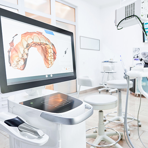
Digital 3D Tooth Impressions – Intraoral Scanner (iTero Intraoral Scanner)
The 3D intraoral scanner can scan a patient’s mouth in real-time, generate 3D images, and has a close-range infrared function that can be used to detect cavities. This helps dentists explain dental issues to patients more easily and improves diagnostic accuracy.
In addition to being a dental examination tool, this advanced 3D scanner can replace traditional plaster impression techniques. The entire impression process only takes 5 minutes, which is 50% faster than traditional techniques, reducing the need for reprinting due to errors, and patients can also reduce discomfort caused by impressions.
Features Of Digital Dental Scanning:
- Real-time simulation of orthodontic results.
- Preview of simulated orthodontic treatment progress and the difficulty of correcting teeth.
- Impression process only takes 5 minutes.
- Fast and comfortable process.
- No radiation involved.
- Does not cause throat sensitivity or reflex vomiting as traditional impressions do.
- Suitable for various dental services including teeth straightening, implants, and braces.


Any medical or healthcare information provided by us on this website is for reference or educational purposes only and we do not warrant that such information will meet your health or medical requirements. Furthermore, our website is NOT intended for use in diagnosing or treating medical or health conditions, and is NOT a substitute for qualified professional medical or healthcare advice and should not be treated as such. You should always seek advice from a qualified medical professional or other healthcare service provider with any questions you may have regarding a medical or health condition.